

Imaging and examination
A panoramic image is used to review the tooth direction, depth, nearby teeth, and surrounding anatomy.
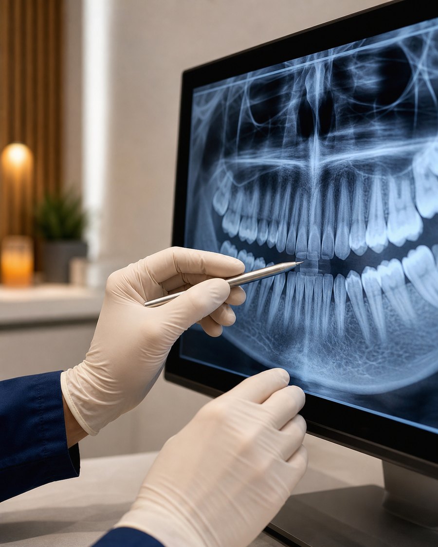
The tooth position and nearby nerve canal are reviewed before the extraction plan is selected.
A wisdom tooth may be monitored when it is healthy, functional, and can be cleaned. Extraction may be considered when it is impacted, repeatedly inflamed, difficult to clean, or affecting the tooth in front.
For lower impacted wisdom teeth, the relationship with the inferior alveolar nerve canal is part of the assessment. Imaging helps determine whether treatment can proceed in the clinic or whether another approach is appropriate.
The choice depends on symptoms, position, and nearby teeth.
3D CT is added when the anatomy needs closer review.
Availability and clinical suitability are confirmed after assessment.
The actual procedure and recovery vary with the tooth position, depth, and individual health.
A panoramic image is used to review the tooth direction, depth, nearby teeth, and surrounding anatomy.
If a lower wisdom tooth appears close to the nerve canal, 3D CT may be used to examine the relationship in more detail.
The clinician explains whether observation, extraction, staged treatment, or referral is appropriate for the findings.
After local anaesthesia and extraction, bleeding control, wound care, medicines, and follow-up are explained.
Contact the clinic if swelling is worsening, you develop fever or difficulty swallowing, bleeding does not settle, pain becomes much worse after first improving, or altered sensation continues. In an emergency, use the appropriate local emergency service.
Clinical suitability is confirmed only after imaging and examination.
Many cases can be assessed and treated on the same day, but this cannot be promised before imaging and examination. The tooth position, nearby nerve canal, medical history, and number of teeth being treated all affect the plan.
A lower wisdom tooth can lie near the inferior alveolar nerve canal. When a panoramic image suggests close proximity, 3D CT may be used to examine the relationship. A staged approach or referral may be recommended when the findings indicate higher risk.
Follow the instructions given after treatment. In general, avoid smoking, drinking through a straw, forceful rinsing, and strenuous exercise during the early recovery period because these can disturb the healing blood clot.
Contact the clinic if swelling continues to increase, fever develops, swallowing becomes difficult, bleeding does not settle, pain becomes markedly worse after initially improving, or altered sensation persists.
Assessment, treatment stages, and planning factors.
Read the guide →Plan your visitAddress, maps, parking, transport, and weekly hours.
View visit information →한국어The full Korean-language clinical guide.
View Korean guide →Please call to confirm assessment availability and any language-support needs before travelling to the clinic.